Paper · Frontiers in Neuroscience · 2020
Wiring the brain straight into a silicon chip
How bundles of glass-coated microwires, pressed straight onto mass-produced amplifier chips, could let brain recording grow at the pace of the chip industry.
Cancer Neurotechnology Lab
14 October 2026 · 10 min read
The chips inside phones and cameras keep getting denser and cheaper. Devices that record from the brain have not kept pace. Recording from more neurons usually means more wires, more connectors and more amplifiers, and each step adds bulk, cost and engineering effort.
A 2020 paper in Frontiers in Neuroscience proposed a different route.[1] Instead of designing a new chip for every new brain-recording experiment, the team took existing, mass-produced amplifier chips and connected them to the brain through bundles of very fine glass-coated wires. They called the approach CHIME, short for CMOS-Hosted in vivo MicroElectrodes.
The scaling problem
Electrical recording is the fastest way to follow brain activity: it captures individual nerve impulses, or spikes, which last about a millisecond. The largest electrical recordings so far rely mainly on silicon probes, thin shanks carrying rows of contacts.
The paper sets out why these are hard to scale.[1] Recording sites on a silicon shank sit along one line, while many well-studied brain areas, including the cortex, hippocampus and olfactory bulb, are organised in horizontal layers that are best sampled across a broad area. Packing many shanks close together damages tissue. And each new custom chip design takes a long time to verify and manufacture.
Flat recording chips built with CMOS technology, the standard process for modern microchips, have already scaled to tens of thousands of channels for brain slices and cell cultures. But those chips are flat, and a living brain is not. CHIME was designed to add the missing third dimension.
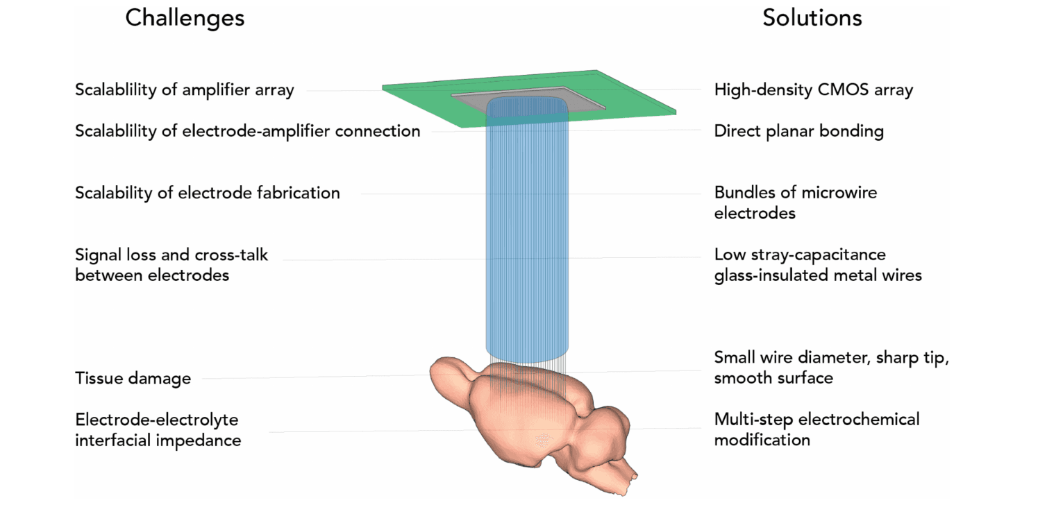
The design idea. Each challenge of scaling up electrical recording (left) is paired with a design choice in CHIME (right): a high-density CMOS chip for amplification, direct contact between wires and chip, bundled microwires, glass insulation, small sharp wires, and a chemically modified tip.
Reproduced from Kollo, Racz, Hanna, Obaid et al., Front. Neurosci. 14:834 (2020), under CC BY.Wires made by the kilometre
The wires are produced by a method in which molten metal is drawn inside glass into a continuous thread.[1] The result is a flexible fibre with a conducting metal core and a smooth glass coat, made in lengths of hundreds of metres. For the recordings in this paper, the fibres measured 22–25 micrometres across, with metal cores of 1–7 micrometres.
Because the fibres are flexible, they can be wound into bundles by machine, much like thread on a spool. The team made bundles of 100 to 1,000 wires. The bundles were set in hard plastic and the two ends treated differently:
- The chip end was polished flat, and small hemispherical gold bumps were grown on each exposed metal core by electrodeposition. Bigger contacts make a reliable connection more likely when the bundle is pressed against the chip.
- The tissue end was polished at a 30-degree angle to give each wire a sharp tip, then freed from its embedding so the last couple of millimetres of each wire could move independently. The tips were given an electrochemical coating to improve recording, as described in the related jULIEs work.[2]
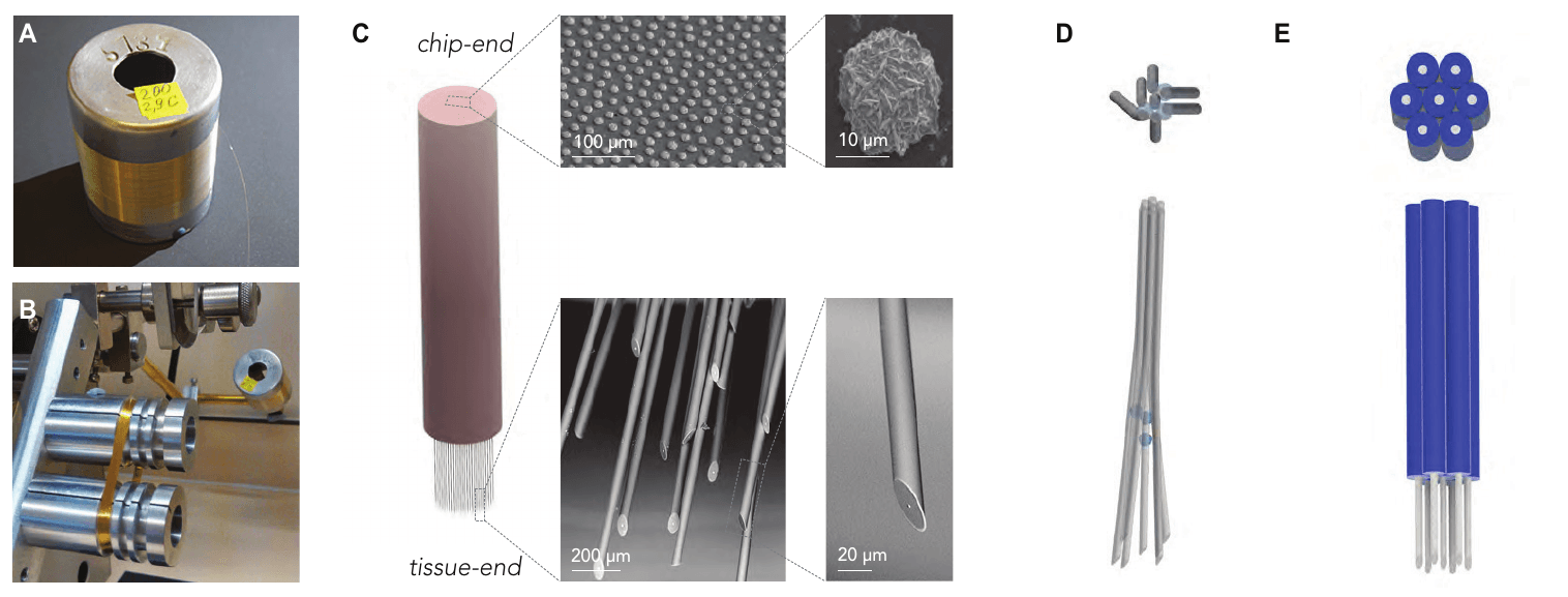
From spool to bundle. Microwire comes on long spools (A) and is wound into bundles (B). The chip end is polished and given gold contact bumps, while the tissue end is sharpened (C). A temporary spacer coating can set the distance between wires (D).
Reproduced from Kollo, Racz, Hanna, Obaid et al., Front. Neurosci. 14:834 (2020), under CC BY.Pressing a bundle onto a chip
The key idea is to skip individual wiring. The polished chip end of the bundle is simply pressed onto the grid of amplifier pixels.[1] Because the chip has far more pixels than the bundle has wires, and the wires are spaced more than 100 micrometres apart, each wire is likely to land on at least one pixel without any careful alignment. Pixels that are touching a wire can be identified afterwards from their signals.
The team tried this with two very different chips:
- A research MEA chip (MEA1k) with 26,400 recording pixels, of which 1,024 can be selected and recorded at once. It allows recording only from the pixels connected to wires.
- The readout chip of an infrared camera (Xenics Cheetah 640CL), supplied without its light-sensitive layer. It contains 327,680 amplifiers and records frames at up to 200 kHz for a small window of pixels, or 1.7 kHz for the full frame. The brain recording is captured as a video stream, and voltage traces are extracted from the connected pixels afterwards.
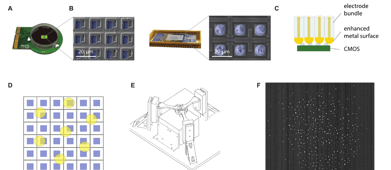
Two chips, one approach. A research MEA chip (A) and an infrared camera readout chip (B) were both connected by pressing the bundle directly onto the pixels (C). With enough spacing between wires, contacts form without precise alignment (D). A three-arm press holds the parts together (E). Bright pixels show where 200 wires made contact with the camera chip (F).
Reproduced from Kollo, Racz, Hanna, Obaid et al., Front. Neurosci. 14:834 (2020), under CC BY.How clean is the signal?
A long, thin wire can lose much of a small signal along the way. The paper measured how much electrical coupling leaks from the wire into the surrounding fluid, and found it very low, under 80 femtofarads per millimetre of wire, because the glass coat is thick compared with the metal core.[1]
Measured noise in saline was 24.2 ± 7.7 microvolts (RMS) on the MEA chip and 58.2 ± 21.5 microvolts on the camera chip. For spikes of 150–500 microvolts, which the authors recorded reliably in the mouse brain, that gives signal-to-noise ratios of 6–20 and 2.5–9 respectively. After removing noise shared across channels, the remaining noise on the MEA chip fell to 6.5 ± 2.6 microvolts, which the authors suggest means it could be reduced further with shielding.
26,400
Pixels on the MEA1k chip (1,024 recordable at once)
327,680
Amplifiers on the camera readout chip
156 ± 36
Connected wires, 200-wire bundles (n = 7)
under 80 fF/mm
Stray capacitance of the glass-coated wire
Recording in the brain
The team recorded from the olfactory bulb, the brain's first relay for smell, in anaesthetised mice.[1] With the MEA chip and 200-wire bundles, an average of 156 ± 36 pixels were connected with good signal quality across seven experiments. Recording started immediately after the bundle was inserted. Sessions lasted from 23 minutes to 2 hours.
Both chips recorded spikes from individual neurons. The camera chip picked up smaller slow waves (local field potentials) because of how its amplifiers filter the signal, but still resolved well-separated single-neuron spikes. In one experiment, 86 channels on the MEA chip recorded spikes around a one-second odour stimulus.
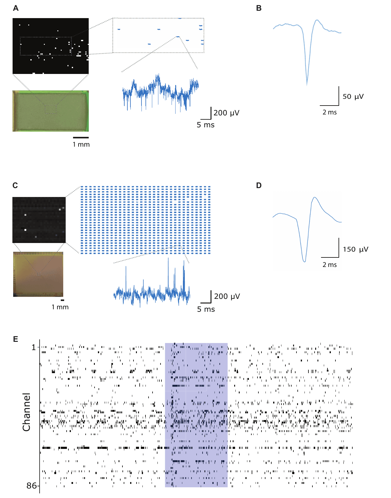
Neurons recorded through both chips. Traces and spike shapes from the olfactory bulb recorded with the MEA chip (A, B) and the camera chip (C, D). Panel E shows spikes on 86 channels, with the timing of a one-second odour stimulus shaded.
Reproduced from Kollo, Racz, Hanna, Obaid et al., Front. Neurosci. 14:834 (2020), under CC BY. This is Figure 6 in the paper.The wires entered the cortex without buckling, consistent with the authors' estimate that a one-millimetre length of this wire can carry a force of about 300 micronewtons before it bends. The bundles were also imaged inside the skull by X-ray computed tomography, where individual wires could be seen, so recording sites can be located against the anatomy.
Damage was discussed with reference to the related studies. The authors note that, although the wires were inserted quickly (at 100 micrometres per second or faster), signals were stable from the start of recording, rather than showing the dip in signal quality often reported during the first 40–60 minutes after an electrode is inserted, which is generally attributed to local tissue disturbance.
What this does and does not show
- What was demonstrated. Glass-coated microwire bundles can be connected directly to two different off-the-shelf CMOS amplifier chips and used to record single-neuron activity in the mouse brain, with most wires making a working connection.[1]
- What was not tested. All recordings were acute and head-fixed. The authors write that recordings stayed stable for over 40 minutes, but that chronic recordings, lasting days to months, will require further histological and functional studies.
- What it is. CHIME is a method and an architecture, not a finished device. The paper presents it as a way to link neural recording to the rapid progress of the chip industry.
Why it matters
For brain–computer interfaces and large-scale neuroscience, the attraction is modularity. The electrode bundle and the electronics are made separately and joined by pressure, so either part can be improved or swapped without redesigning the other. The authors note that the same approach could be extended to other CMOS arrays, such as chips that deliver electrical stimulation.
A companion paper in Science Advances, from the same collaboration, scaled the bundles to thousands of wires and recorded from awake, moving mice.[3] Read our guide to that paper.
Who did the work
Mihaly Kollo, Romeo Racz, Mina-Elraheb Hanna and Abdulmalik Obaid contributed equally as joint first authors, with Matthew R. Angle, William Wray, Yifan Kong, Jan Müller and Andreas Hierlemann. Nicholas A. Melosh and Andreas T. Schaefer share senior authorship. The work brought together the Francis Crick Institute and University College London, Stanford University, Paradromics Inc., ETH Zürich and MaxWell Biosystems.[1]
According to the paper's author contributions, Kollo, Racz, Hanna, Obaid, Angle, Schaefer and Melosh initiated the concept and design; Kollo, Racz and Wray performed the experiments, data acquisition and analysis.
Competing interests, as declared in the paper. Several authors are employed by and/or hold shares in Paradromics, Inc., a brain–computer interface company, and one author is a co-founder of MaxWell Biosystems AG. The manuscript was first released as a preprint on bioRxiv.
All animal experiments were approved by the local ethics panel and the UK Home Office under the Animals (Scientific Procedures) Act 1986, as stated in the paper.
Sources
- [1]Kollo M, Rácz RR, Hanna M-E, et al. CHIME: CMOS-hosted in vivo microelectrodes for massively scalable neuronal recordings. Frontiers in Neuroscience 14:834 (2020). Open access. doi.org/10.3389/fnins.2020.00834
- [2]Rácz RR, Kollo M, et al. jULIEs: nanostructured polytrodes for low traumatic extracellular recordings and stimulation in the mammalian brain. Journal of Neural Engineering 19(1):016041 (2022). Open access. doi.org/10.1088/1741-2552/ac514f
- [3]Obaid A, Hanna M-E, Wu Y-W, Kollo M, Rácz R, et al. Massively parallel microwire arrays integrated with CMOS chips for neural recording. Science Advances 6(12):eaay2789 (2020). Open access. doi.org/10.1126/sciadv.aay2789
How to cite the original work: Kollo M, Racz R, Hanna M-E, Obaid A, Angle MR, Wray W, Kong Y, Müller J, Hierlemann A, Melosh NA, Schaefer AT. CHIME: CMOS-hosted in vivo microelectrodes for massively scalable neuronal recordings. Frontiers in Neuroscience. 2020;14:834. doi:10.3389/fnins.2020.00834
Figures are reproduced from the original open-access article under the CC BY licence. Captions have been rewritten for a general audience; see the paper for the authors’ full captions.
This summary is written for public understanding and is not medical advice.